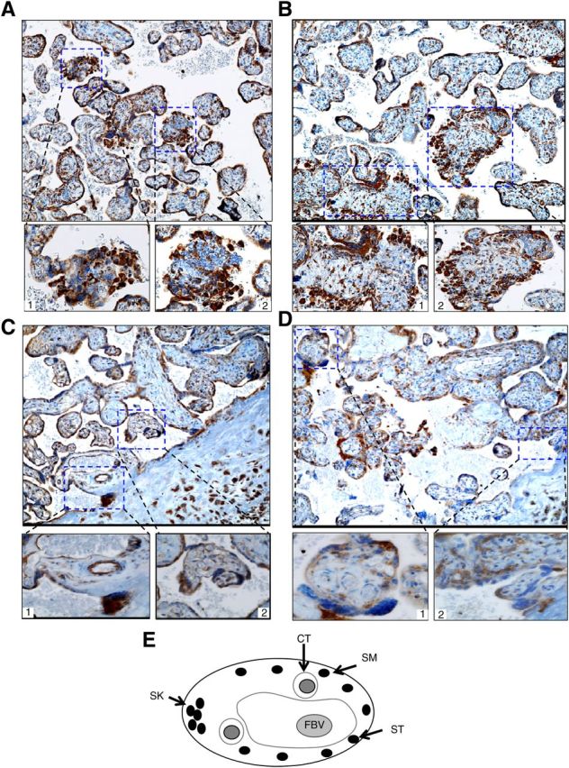
Fig. 2.

Immunohistochemical analyses of NF-κB components and CRH in human term placenta. Four sections from the same placenta were stained with antibody to human RelB (A), NF-κB2 (B), CRH (C), or Rel A (p65) (D), respectively. Representative images are shown in the top panel; original magnification, ×100. Bottom panels represent insets; original magnification, ×400. E, Schematic presentation of cross-section of human term placental villus. CT, Cytotrophoblasts; ST, syncytiotrophoblast; SM, syncytium; SK, syncytial knot; FBV, fetal blood vessel.
