Fig. 3.
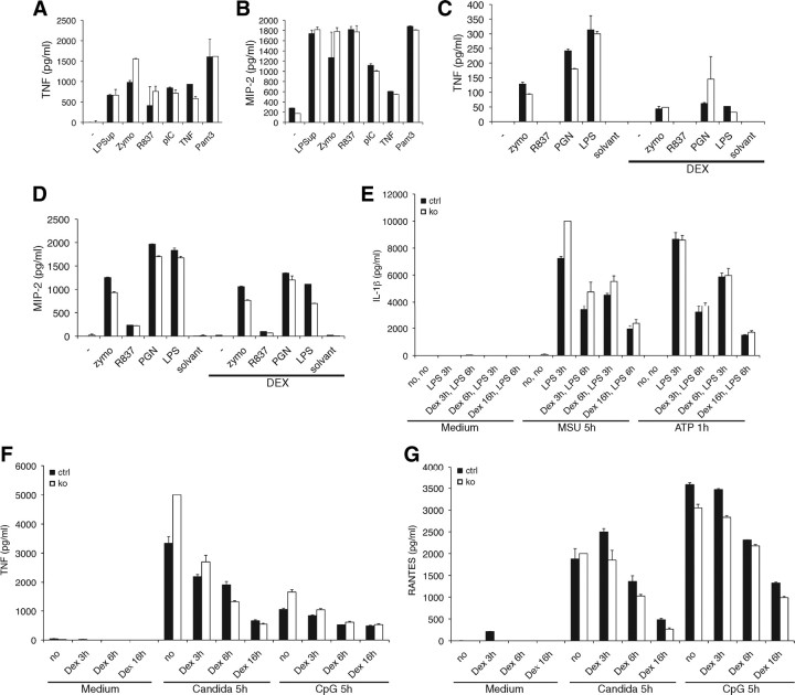
Tsc22d3-2Δ/y mice show no impairment of inflammatory responses. Peritoneal and bone marrow-derived cytokine secretion. (A and B), ELISA for TNF (A and C), and MIP-2 (B and D) after stimulation or not of peritoneal macrophages with LPS supplemented by indicated stimuli. C and D, Stimulation of BMDM (see A and B) with or without dexamethasone (DEX) (0.2 mm). Data are shown as mean values of duplicated stimulations ± sem from three mice per group. E–G, Quantification of IL-1β (E), TNF and Rantes (G) production in bone marrow-derived cells primed with 10 ng/ml LPS with or without dexamethasone (Dex) for indicated periods before inflammasome activation by 100 ng/ml MSU for 5 h or 5 mm ATP for 1 h) (E), heat-inactivated Candida cells (F) or 1 μm CpG for 6 h (G). Data are shown as mean values of triplet stimulations ± sem and representative for three independent experiments with a total of six control and six knockout mice.

