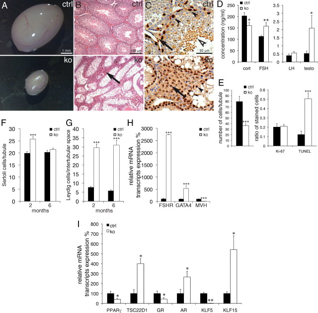
Fig. 5.
Tsc22d3-2 knockout males are sterile. A, Representative pictures from 2- and 6-month-old control (top) and knockout (bottom) adult testes; B, H&E-stained paraffin sections; C, immunohistochemistry of Sertoli and Leydig cells counterstained with H&E-stained paraffin sections from control and knockout testes. In B and C, note absence of mature spermatozoa (white arrowhead) and apparent hyperproliferation of Leydig cells (black arrow) in the knockouts. Panel C is a higher-magnification image (rabbit anti-GATA4 antibody) with counterstaining with hematoxylin (blue nuclei); elongated spermatids (white arrowhead) are seen in controls. D, Corticosterone (cort), FSH, LH, and testosterone (testo) hormone levels in plasma (n ≥ 10 animals per group, 3–4 months old). E, Total number of cells per seminiferous tubule (n ≥ 20 tubules per group; left), ratio of Ki-67-positive cells divided by total cell number, middle), and ratio of TUNEL-positive cells divided by total cell number per seminiferous tubule, n = 18 tubules, right). F, Quantification of Sertoli cells per tubule (F) (n ≥ 25 tubules) and Leydig cells in the intertubular space (G) (n ≥ 27) at 2 months (n = 2) and 6 months (n = 4 animals). H, qRT-PCR of Fshr, Gata4, and Mvh control (black bars) and knockout (white bars) animals. I, qRT-PCR of Pparγ2, Tsc22d1, Gr, Ar, Klf5, and Klf15 in testis from control (black bars) and knockout (white bars) animals. Scale bar, 1 mm (A), 100 μm (B), and 50 μm (C). *, P < 0.05; **, P < 0.01; ***, P < 0.001. ctrl, Control; ko, knockout.

