Fig. 1.
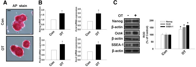
Effect of OT on undifferentiated marker genes of mouse ESC. A, AP enzyme activity was assessed in mouse ESC treated in the presence or absence of OT (10−7 m) for 72 h as described in Materials and Methods. Scale bars, 20 μm (magnification ×400). B, The cells were treated with OT (10−7 m) for 72 h. Total RNA from mouse ESC was reverse transcribed, and Nanog, Oct4, FoxD3, Sox2, and β-actin cDNA were amplified by real-time PCR as described in Materials and Methods. The data are reported as the mean ± se of four independent experiments, each conducted in triplicate. *, P < 0.05 vs. control. C, The cells were treated with OT (10−7 m) for 72 h. Total protein was extracted and blotted with Nanog, Oct4, SSEA-1, and β-actin antibodies. Each example shown is representative of four independent experiments. The right part (C) depicting the bars denotes the mean ± se of four independent experiments for each condition determined from densitometry relative to β-actin. *, P < 0.05 vs. control. Con, Control; ROD, Relative Optical Density.

