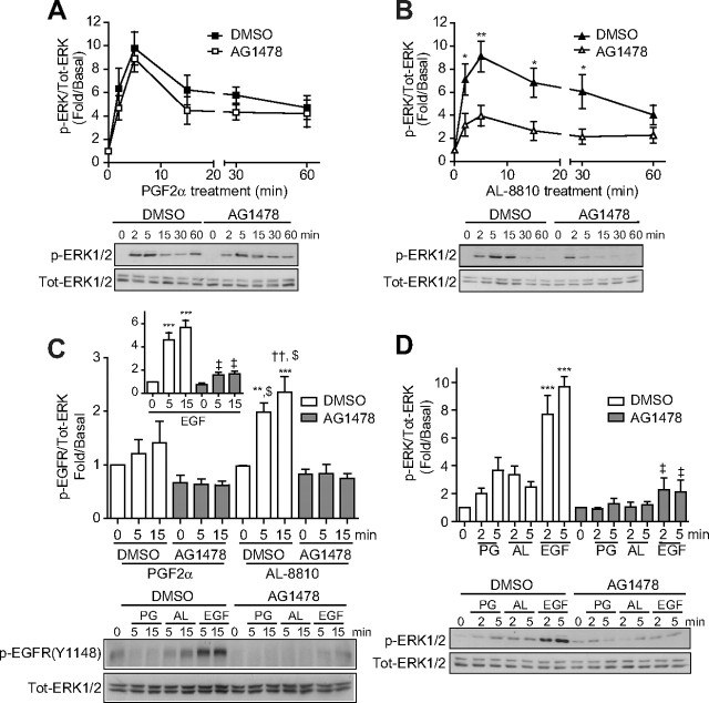
Fig. 5.
AL-8810 activates MAPK through EGFR-dependent transactivation. A–D, FP-R cells (A, B, and C) and MG-63 osteoblasts cells (D) were serum starved for 30 min (A and B) or overnight (C and D) before pretreatment with either DMSO or 125 nm AG1478 for 30 min. Cells were then treated with 1 μm PGF2α (A, C, and D), 10 μm AL-8810 (B, C, and D) or 5 ng/ml EGF (C and D) for the indicated times. Cell lysates were analyzed by Western blot using antiphospho-ERK1/2, antitotal-ERK1/2 (A, B, and D) and anti-phospho EGFR (C) antibodies. Signals were quantified by densitometry and plotted as fold over basal (i.e. not treated). Results are representative of three (D), five (A and B), or six (C) independent experiments. *, P < 0.05; **, P < 0.01, comparing DMSO and AG1478 treatment for each corresponding time points of ligand stimulation (panels A and B); **, P < 0.01; ***, P < 0.0001 compared with DMSO-treated, ligand-unstimulated cells (panels C and D); ††, P < 0.01 compared with DMSO-pretreated, PGF2α-stimulated cells, at 5 and 15 min; $, P < 0.0001 compared with AG1478-pretreated and PGF2α- and AL8810-stimulated cells (panel C); ‡, P < 0.0001 compared with DMSO-pretreated, EGF-stimulated cells (panel C, inset, and panel D). AL, AL-8810; Tot, total.

