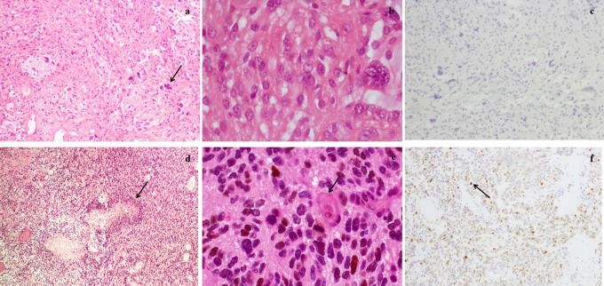
Figure 1.
Histopathology: (A) Surgical specimens from the first operation stained with hematoxylin-eosin (×100), demonstrating the fasciculate pattern, mucoid regions, and bipolar neoplastic cells and some giant cells (arrow). B, Stained with hematoxylin-eosin (×400), absence of mitosis and cells with nuclear anaplasia. C, Negative representative results of p53 immunohistochemical staining. D, Surgical specimens from the second operation stained with hematoxylin-eosin (×100), showing hypercellularity, with small and anaplastic cells and necrosis with a palisade pattern (arrow). E, Stained with hematoxylin-eosin (×400), with mitosis (arrow) and endothelial proliferation. F, Positive representative results of p53 immunohistochemical staining (arrow).

