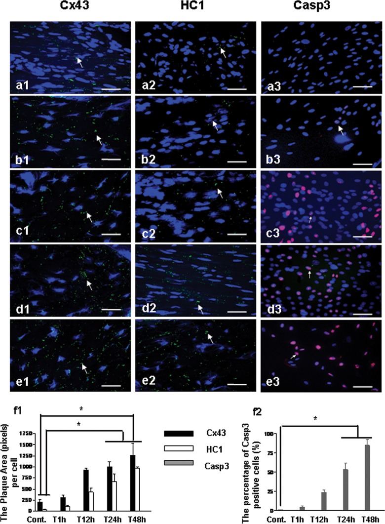
Figure 2.
Immunostaining for the expression of Cx43, HC1, and Casp3 in rat’s brain after HI insult. Cx43, HC1, and Casp3 were stained green, and nuclei were stained blue. Bar = 25 µm. a1-3, The expression of Cx43, HC1, and Casp3 in SVZ from rat without the insult of HI (Cont.); b1-3, The expression of Cx43, HC1, and Casp3 in SVZ from rat 1 hour after HI (T1 hour); c1-3, The expression of Cx43, HC1, and Casp3 in SVZ from rat 12 hours after HI (T12 hour); d1-3, The expression of Cx43, HC1, and Casp3 from rat 24 hours after HI (T24 hour); e1-3, The expression of Cx43, HC1, and Casp3 in SVZ from rat 48 hours after HI (T48 hour); f1, The plaque area (pixels) of Cx43 and HC1 staining per cell with the time points. f2, The percentage of Casp3 positive cells (%) with the time points. *P < .05. Cx43 indicates connexin 43; Casp3, caspase 3; HCl, hemichannel; HI, hypoxia–ischemia; SVZ, subventricular zone.

