
Figure 1.
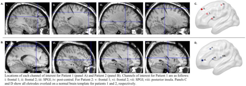
Locations of each channel of interest for Patient 1 (panel A) and Patient 2 (panel B). Channels of interest for Patient 1 are as follows: i: frontal 1; ii: frontal 2; iii: SPGI; iv: post-central. For Patient 2: v: frontal 1; vi: frontal 2; vii: SPGI; viii: posterior insula. Panels C and D show all electrodes overlaid on a normal brain template for Patients 1 and 2, respectively.