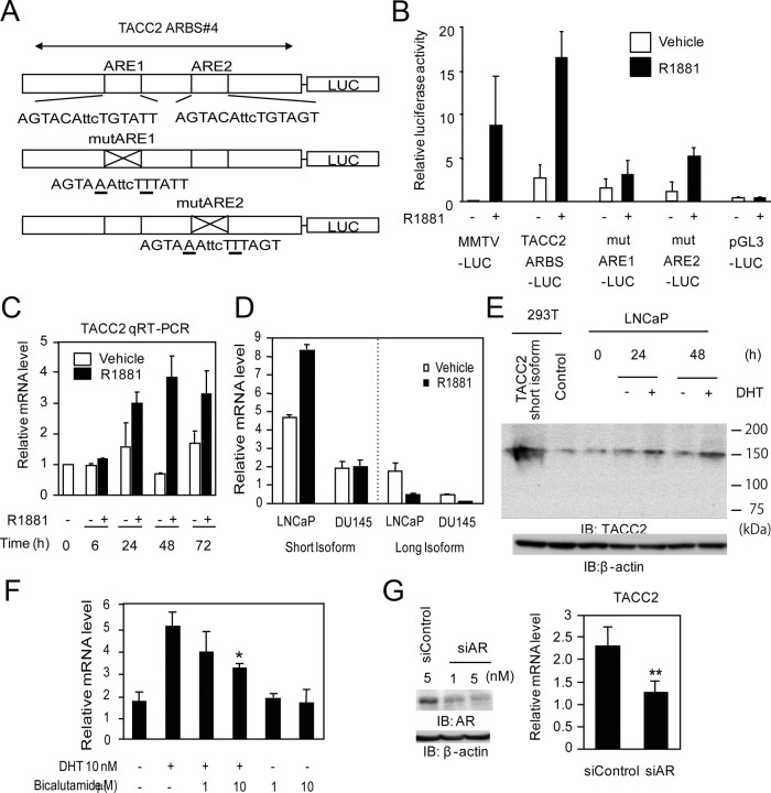
Fig. 2.
AR- and ligand-dependent transcription of TACC2. A, Schematic view of luciferase reporter constructs of TACC2 ARBS 4 (TACC2 ARBS-LUC: containing intact ARE, mutARE1-LUC: mutated ARE1 with intact ARE2, and mutARE2-LUC: intact ARE1 with mutated ARE2). Mutated bases in ARE are underlined. B, Luciferase activity of TACC2 ARBS luciferase vectors transfected into LNCaP cells. The pGL3 promoter and mouse mammary tumor virus (MMTV)-LUC vectors were used as negative and positive controls, respectively. Cells were treated with R1881 (10 nm) or vehicle for 24 h. The data represent the mean ± sd, n = 3. C, Androgen-dependent induction of TACC2 mRNA in LNCaP cells. Cells were stimulated with R1881 (10 nm) or vehicle and subjected to total RNA extraction for 6–72 h. The TACC2 mRNA level was analyzed by real-time PCR. The data represent the mean ± sd, n = 2. D, Isoform-specific regulation of TACC2 by androgen in prostate cancer cell lines. LNCaP cells or DU145 cells were stimulated with R1881 (10 nm) or vehicle and subjected to total RNA extraction for 24 h. TACC2 short or long isoform mRNA levels were analyzed by real-time PCR using specific primers. The data represent the mean ± sd, n = 2. E, Androgen-dependent TACC2 protein induction in LNCaP cells. Cells were treated with DHT (10 nm) or vehicle for 24 and 48 h. Whole-cell lysates were immunoblotted by anti-TACC2 and anti-β-actin. 293T cell lysates transfected with the expression vector for a short form of TACC2 (NM_206860) or empty vector were used as positive or negative controls, respectively. F, Modulation of TACC2 mRNA levels by DHT and antiandrogen bicalutamide. LNCaP cells were treated with vehicle, DHT (10 nm), DHT (10 nm) plus bicalutamide (1 or 10 μm), or bicalutamide (1 or 10 μm) alone for 24 h, and subjected to total RNA extraction. The TACC2 mRNA level was analyzed by real-time PCR. The data represent the mean ± sd, n = 3. *, P < 0.05 from DHT alone. G, Knockdown of AR represses TACC2 transcription. LNCaP cells were transfected with siControl (5 nm) or siAR (1 nm or 5 nm). G (left panel), Whole-cell lysates were immunoblotted with anti-AR 48 h after transfection. Anti-β-actin was used as a loading control. G (right panel), LNCaP cells were transfected with siControl (5 nm) or siAR (5 nm). The TACC2 mRNA level was analyzed by real-time PCR. The data represent the mean ± sd, n = 3. **, P < 0.01 from siControl. IB, Immunoblotting; mut, mutant; qRT-PCR, quantitative RT-PCR..

