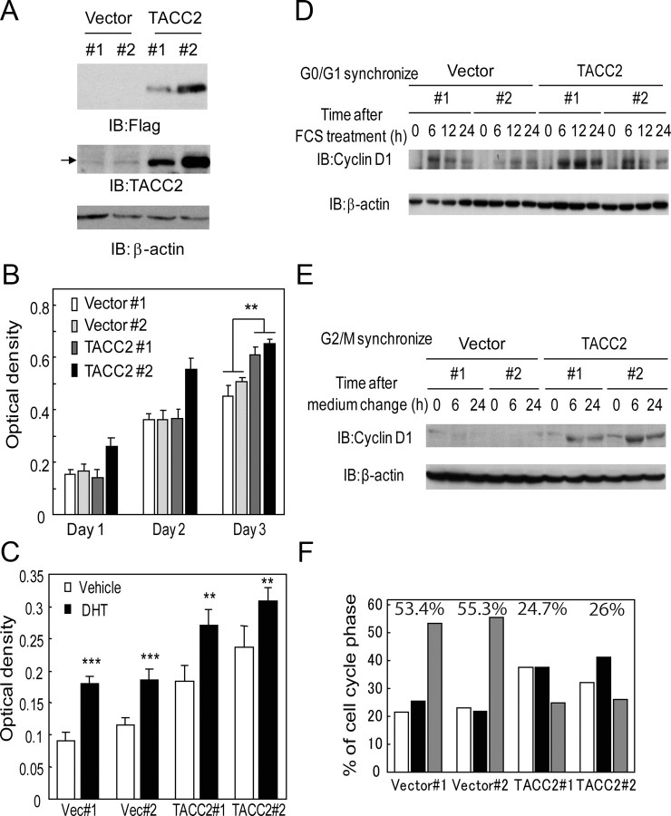
Fig. 6.
TACC2 overexpression accelerates the cell cycle at G2/M phase for prostate cancer cell proliferation. A, Generation of LNCaP cells stably expressing TACC2. Expression of exogenous FLAG-tagged TACC2 was confirmed by Western blotting using anti-FLAG and anti-TACC2 antibodies. β-Actin antibody was used as a loading control. Arrow indicates the bands of endogenous TACC2 protein. B, Ectopic TACC2 expression promotes cultured LNCaP cell growth. Cell viability was measured by the MTS assay. The data represent the mean ± sd, n = 4. **, P < 0.0001 from vector-expressing cells (Vector 1 and 2). C, Androgen-dependent cell proliferation in TACC2 overexpressing LNCaP cells. Vector-expressing and TACC2-expressing LNCaP cells were treated with 10 nm DHT or vehicle. Cell viability was measured 3 d after treatment by the MTS assay. The data represent the mean ± sd, n = 4. **, P < 0.001; ***, P < 0.0001 from vehicle-treated cells. D and E, TACC2 overexpression accelerates cell cycle progression after G2/M synchronization. D, After serum starvation for 24 h, vector-expressing or TACC2-overexpressing cells were treated with 20% FCS for 0, 6, 12, and 24 h, after which cell lysates were immunoblotted with anti-cyclin D1 and β-actin antibodies. E, After treatment with nocodazole (0.5 μg/ml) for 14 h, plates were washed and cells were incubated in fresh medium. Cell lysates after 0-, 6-, and 24-h incubation were immunoblotted with anticyclin D1 and anti-β-actin antibodies. F, TACC2 overexpression promotes the cell cycle of LNCaP cells after G2/M synchronization. After cells were treated with nocodazole (0.5 μg/ml) for 14 h, the medium was changed to fresh medium. Cell cycle analysis was performed after a 24-h incubation period. Representative result of repeated three experiments (two clones for each group) is shown. Percentage of G2/M phase cells for each clone is described in the graph. IB, Immunoblotting;

