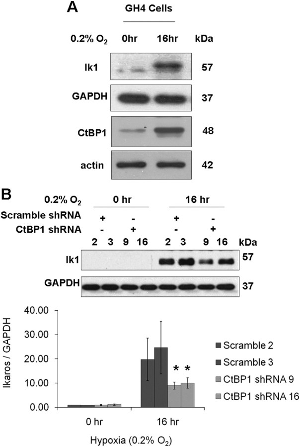
Fig. 4.
Hypoxia enhances the expression of Ikaros in pituitary tumor cells, and CtBP1 deficiency diminishes this effect. A, Western blotting depicting CtBP1 levels and Ikaros in wild-type GH4 cells before and after the indicated 16 h of hypoxia. Neither actin nor GAPDH loading controls are affected by hypoxia in these cells. B, Western blotting depicting Ikaros levels in control and CtBP1-deficient GH4 cells before and after 16 h of hypoxia. GAPDH was detected as a loading control. Densitometric quantification of Ikaros expression is normalized to GAPDH. Bar graphs indicate the mean values of two independent experiments, and sd are depicted by error bars. *, Significance according to the Student's paired t test: P < 0.01.

