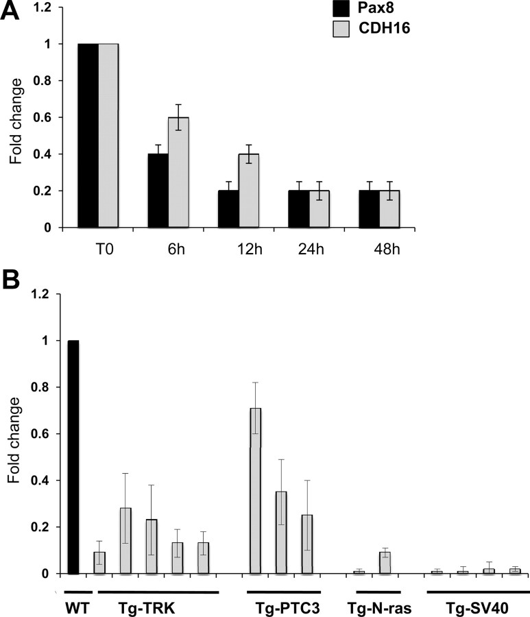
Fig. 7.
Cadherin-16 expression is down-regulated in dedifferentiated thyroid cells. A, qRT-PCR analysis was performed on total RNA prepared from FRTL-5 and FRTL-5/ERTM-RAS cells. The expression of Pax8 (black bars) and Cadherin-16 (CDH16) (gray bars) mRNA was measured after a time course of 4OHT treatment (6, 12, 24, and 48 h). The values are means ± sd of three independent experiments in duplicate, normalized by the expression of β-actin, and expressed as fold change with respect to the T0, whose value was set at 1.0. Statistical analysis uses t test (P < 0.01). B, qRT-PCR analysis was performed on total RNA prepared from thyroid carcinomas developed in Tg-TRK, Tg-PTC3, and Tg-N-ras transgenic mice expressing the TRK, RET/PTC3, and N-ras oncogenes under the transcriptional control of the Tg promoter. Relative quantities of Cadherin-16 mRNA were normalized to the cyclophilin-A as reference gene. Each amplification was performed in duplicate, and the data are expressed as fold change with respect to the normal mouse thyroid tissues used as a control (WT), whose value was set at 1.0. Statistical analysis uses t test (P < 0.01).

