
Fig. 1.
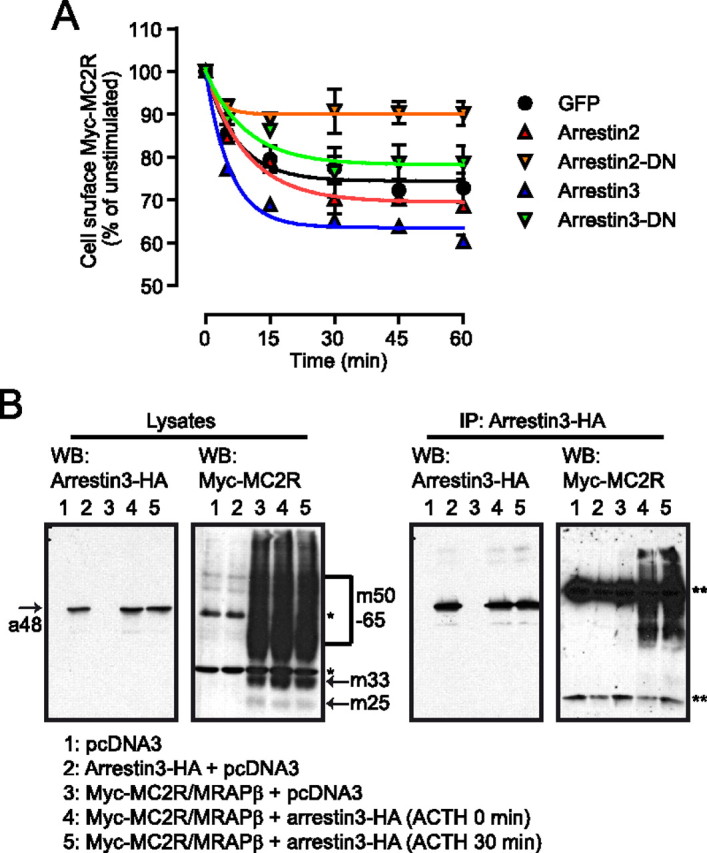
Interaction between arrestins and MC2R. A, Cells were transiently transfected with either control vector (GFP), Arr2, Arr2-DN, Arr3, or Arr3-DN and stimulated with ACTH 100 nm for the selected time periods after which cell-surface Myc-MC2R was measured by ELISA (n = 3). Data points were fitted with a decaying function. B, Native HEK293 cells were transiently cotransfected with pcDNA3, HA-Arr3, and Myc-MC2R/MRAPβ-Flag vectors as indicated. HA-Arr3 was immunoprecipitated and the resulting immunoprecipitates, and input lysates, were analyzed by reducing SDS-PAGE. Western blotting (WB) analyses against the HA and Myc tags are illustrated. a48 indicates Arr3 band, 48 kDa; m25, m33, and m50-65 designate the native, core-, and terminally glycosylated forms of MC2R, respectively; *, endogenous c-Myc (60 kDa); **, IgG light and heavy chains in the anti-Myc immunoblot after immunoprecipitation (IP).