
Fig. 2.
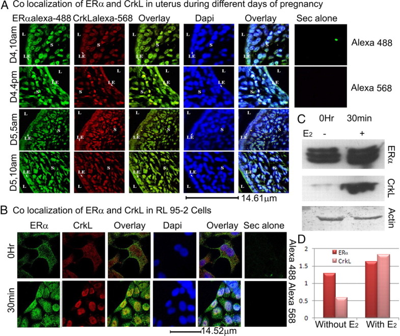
ERα and CrkL colocalize. A, Colocalization of ERα and CrkL in the uterine sections. Paraffin-embedded uterine sections of different days of pregnancy: preimplantation (d 4, 1000 h), late preimplantation (d 4, 1600 h), periimplantation (d 5, 0500 h), and postimplantation (d 5, 1000 h) periods were used. ERα probed with the primary antibody (sc-542) was labeled with antirabbit secondary Alexa Fluor 488, and CrkL probed with primary (sc-319) was labeled with Alexa Fluor 568. DAPI was used as the nuclear stain. Images were taken using the Leica TCS SP2 laser scanning confocal microscope. B, Colocalization of ERα and CrkL in RL95-2 cell line. Secondary alone controls for both Alexa Fluor 488 and Alexa Fluor 568 are shown. C, Western blot analyses of ERα and CrkL using the RL95-2 total cell extracts before and after estrogen treatment and its associated histogram (D). Actin is used as loading control.