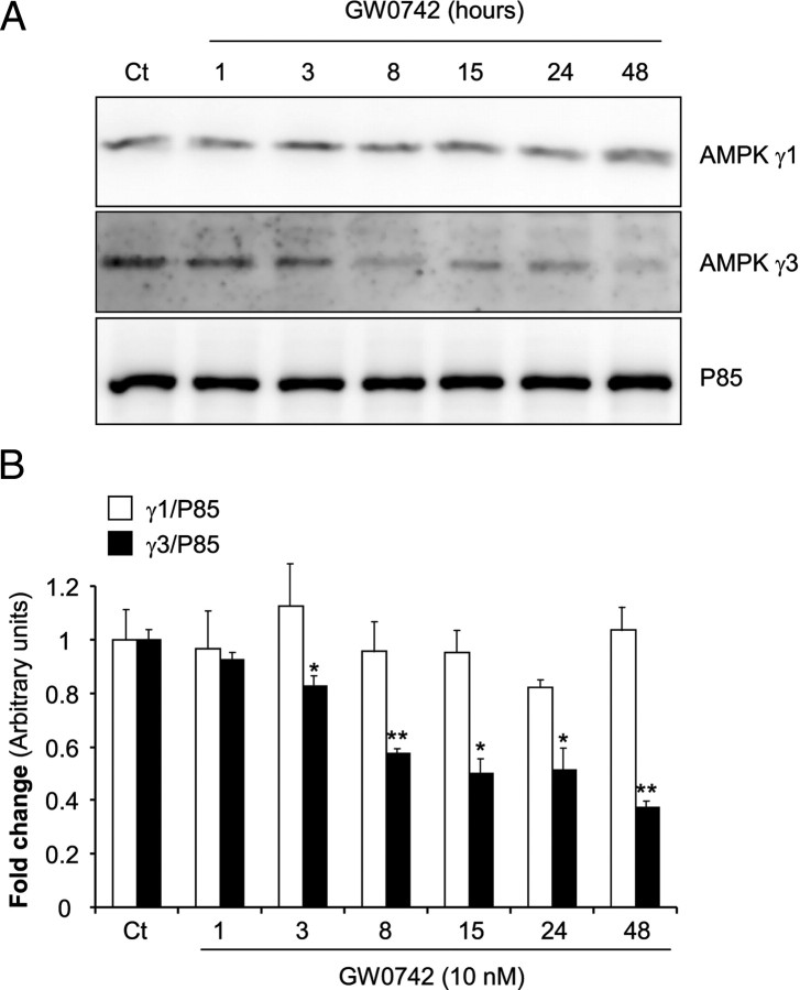
Fig. 7.
PPARβ activation leads to rapid reduction of γ3-AMPK protein levels in C2C12-PPARβ myotubes. a, Representative Western blot analyses of γ1-AMPK, γ3-AMPK, and P85 (loading control) using protein lysates from C2C12-PPARβ cells treated with GW0742 (10 nm) for increasing periods of time. b, After densitometric analysis, results were normalized against the loading control (P85), pooled, and represented as histograms. Values are means ± sd. *, P < 0.05; **, P < 0.005 when compared with the value of the corresponding AMPK subunit in control (Ct) groups (cells treated with vehicle).

