Fig. 4.
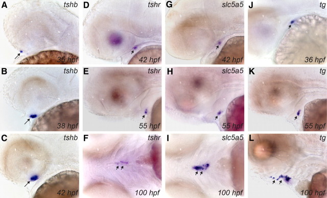
Developmental expression of tshb and thyroid differentiation markers in zebrafish embryos as demonstrated by WISH. Lateral views of the head region show the early expression pattern of tshb in pituitary (A–C) and of tshr (D and E), slc5a5 (G and H), and tg (J and K) in thyroid primordium. Note the earlier onset of tg expression relative to tshr and slc5a5 and the strong staining of the thyroid at the one-follicle stage (55 hpf) by all three thyroid markers. At 100 hpf, dispersed thyroid tissue along the pharyngeal midline is strongly labeled by riboprobes for tshr (ventral view in F), slc5a5 (ventral view in I), and tg (lateral view in L). Long arrows point to pituitary, and short arrows point to thyroid. All embryos are oriented with anterior to the left.

