Fig. 5.

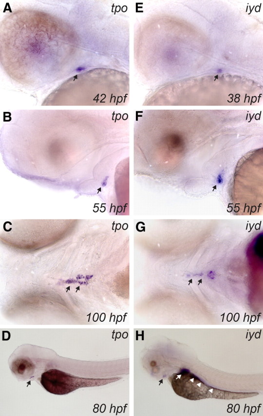
Developmental expression of tpo and iyd in zebrafish embryos as demonstrated by WISH. Lateral views of the head region show the early expression pattern of tpo (A and B) and iyd (E and F) in thyroid primordium. Note the earlier onset of iyd thyroid expression (∼38 hpf) relative to tpo (40–42 hpf) and the strong expression of both genes in the thyroid at the one-follicle stage (55 hpf). Ventral views show that tpo (C) and iyd (G) are strongly expressed in differentiated thyroid tissue at 100 hpf. Black arrows point to thyroid. Although the tpo riboprobe (lateral view in D) specifically stains the thyroid, iyd (lateral view in H) is also strongly expressed in liver (white arrow) and gut (white arrowheads). All embryos are oriented with anterior to the left.
