Fig. 7.
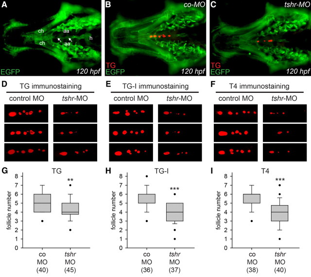
Effects of tshr knockdown on whole-mount immunofluorescence staining for TG, TG-I, and T4 in Tg(fli1a:EGFP) transgenic larvae. A–C, Ventral views of the head region of 120 hpf larvae oriented with anterior to the left. A, The anatomical positions of the ceratohyal (ch), ventral aorta (white arrows), aortic arch arteries (aa), and heart (h) are highlighted by EGFP immunofluorescence. TG immunofluorescence shows the presence of immunoreactive thyroid follicles in embryos injected with control morpholino (co-MO) (B) and tshr morpholino (tshr-MO) (C). D–F, Representative selection of immunostainings (magnified ventral views of thyroid region, anterior to the left) obtained at 120 hpf for TG, TG-I, and T4 in embryos injected with co-MO and tshr-MO. Thyroid tissue of tshr morphants was characterized by a reduced number of functional follicles, many of which displaying a strongly reduced size. Boxplots in G–I show results from quantification of the number of follicles immunoreactive for TG, TG-I, and T4, respectively. Black dots mark outliers and the number of larvae analyzed is indicated in parentheses. Asterisks denote statistically significant differences (Mann-Whitney-test: **, P < 0.01; ***, P < 0.001).

