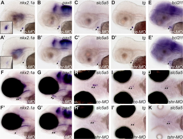
Fig. 9.
tshr morphants display reduced thyroid marker expression as demonstrated by WISH. Lateral views of control embryos (A–E) and tshr morphants (A'–E') analyzed for nkx2.1a, pax8, slc5a5, tg, and bcl2l1 expression at 55 hpf show reduced thyroid expression in tshr morphants. From the staining patterns, timely formation of a first follicle is barely detectable in tshr morphants (see insets in A and A'). Qualitatively similar effects were observed in embryos raised in the presence or absence of 0.003% PTU. For a better visualization of staining in the thyroid region, photomicrographs in A–E and A'–E' show casper larvae raised in PTU. At 100 hpf, analyses of nkx2.1a, pax8, slc5a5, and tg expression in control larvae (F–I) and tshr morphants (F'–I') showed strongly reduced expression of all genes in tshr morphants. Lateral (F, F', G, and G') and ventral views (H, H', I, and I') are shown. Thyroid staining was barely detectable in many tshr morphants. On the other hand, tg staining indicates that the anterior-posterior extension of the pharyngeal midline region populated by thyroid cells is similar between control larvae and tshr morphants (compare I and I'). All photomicrographs in F–I and F'–I' show casper larvae raised in the absence of PTU. Qualitatively similar effects were observed in PTU-treated larvae, but due to enhanced expression of thyroid markers in PTU-treated larvae, the differences in staining intensities were more pronounced. Ventral views in J and K show slc5a5 expression in tshr morphants raised in the absence (J) or presence (K) of PTU. Note that slc5a5 expression was not increased in PTU-treated tshr morphants relative to untreated tshr morphants. All larvae are oriented with anterior to the left. Arrows point to thyroid. co-MO, Embryos injected with standard control morpholino; tshr-MO, embryos injected with tshr morpholino.

