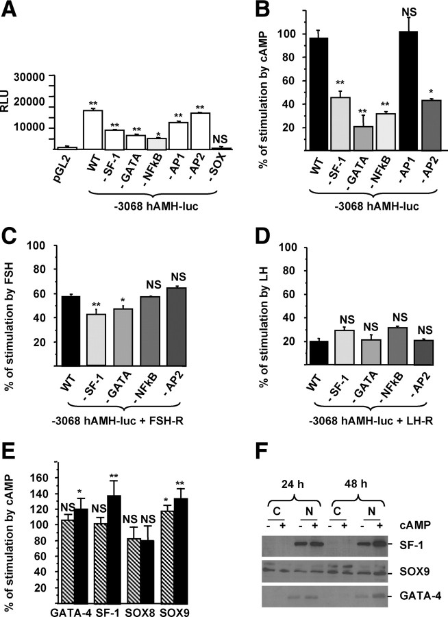
Fig. 6.
Identification of the transcription factors involved in cAMP and gonadotropin stimulation of the AMH gene. A, Cis-activating capacity of the −3068hAMH-luc constructs with mutations in binding sites for NFκB, AP2, GATA-4, SF-1, AP1, and SOX. KK1 cells were transfected with 1 μg of the constructs, and luciferase activity was analyzed after 48 h of culture in control medium. Results are expressed in RLU. B–D, Regulation of the mutant constructs by cAMP (B), FSH (C), and LH (D). KK1 cells were transfected with 1 μg of mutated constructs and 1 μg of FSH-R (C) or LH-R (D), and luciferase activity was assessed 48 h after treatment with cAMP (1 mm) (B), FSH (1 IU/ml) (C), or LH (1 IU/ml) (D). Results are expressed as percentage of stimulation. Data shown correspond to the mean ± sem of three experiments, each done in triplicate. Comparisons of means between different experimental conditions were made by repeated-measures ANOVA, followed by Dunnett post hoc test to compare all vs. control (pGL2 for A and −3068hAMH-luc (WT) for B– D). E, GATA-4, SF-1, SOX9, and SOX8 mRNAs were analyzed by RT-PCR, and their regulation by cAMP in KK1 cells was quantified by real-time RT-PCR after 24 (hatched bars) and 48 (black bars) hours of treatment by cAMP (1 mm). Results are expressed as percentage of response compared with untreated cells using the Student's t test. Data shown correspond to the mean ± sem of three experiments, each done in triplicate. F, Nuclear translocation of SF-1, SOX9, and GATA-4 in the presence of cAMP. SF-1, SOX9, and GATA-4 proteins in cytosol and nuclear fractions (50 μg of each) were analyzed by Western blotting after 24 and 48 h of treatment with cAMP. NS, Not significant; *, P < 0.05; **, P < 0.01.

