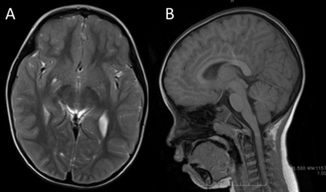
Figure 2.

A, Axial T2-weighted and (B) sagittal T1-weighted brain magnetic resonance imaging of the patient at 4 years of age. Imaging did not reveal any abnormalities except for perivascular space in the lower part of the right basal ganglia. Cerebellar anomaly and atrophy were not observed.
