
Fig. 1.
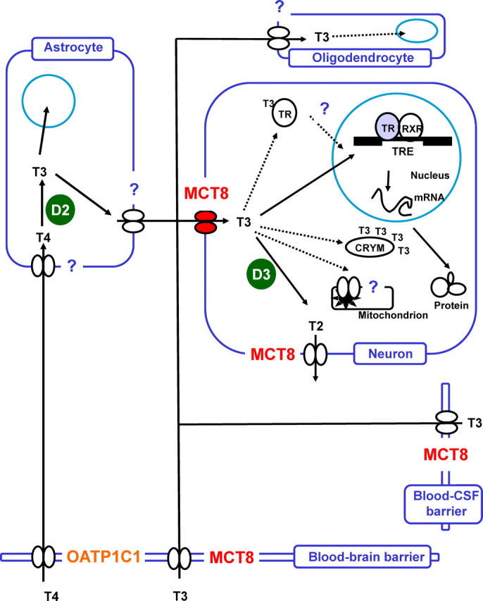
Model of TH regulation in brain. Transporters (paired ovals) are required for uptake and release of T4 and T3. D2 converts T4 to bioactive T3, whereas D3 degrades T3 to 3,3′-T2. T3 in brain is either derived from the circulation [via the blood-brain barrier or indirectly via the blood-cerebrospinal fluid (CSF) barrier] or locally produced from T4 in astrocytes by D2. Intracellular T3-binding proteins (e.g. CRYM) may store cytoplasmic T3 and function in intracellular T3 transport. Ultimately, T3 binds to its nuclear receptor (TR) and modulates gene expression of T3-target genes. T3 actions in mitochondria also require transporter proteins. All principal cells of the brain are T3 targets: neurons, astrocytes, and oligodendrocytes. The degree of certainty of this model is reflected by the color of the symbols. Filled symbols reflect established knowledge (the knowns). White symbols are surmised concepts (the unknowns). Question marks indicate possible novel players or functions in local TH regulation (the guesses). RXR, Retinoic X receptor; TRE, T3-responsive element.