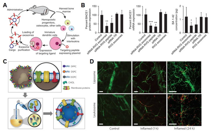
Fig. 6. Natural membrane vesicles.
A) Schematic demonstrating workflow for the fabrication and subsequent administration of siRNA-loaded exosomes for brain-targeted delivery. B) Quantification of BACE1 protein and mRNA expression as well as β-amyloid concentration demonstrates significant knockdown efficacy for the nanoformulation. C) Schematic demonstrating the fabrication of leukosomes via the isolation of membrane proteins followed by reconstitution with synthetic lipids. D) Intravital microscopy images demonstrate preferential accumulation at the site of inflamed tissue over time. Scale bars = 50 μm. A, B adapted with permission from [131]. Copyright Nature Publishing Group, 2011. C, D adapted with permission from [150]. Copyright Nature Publishing Group, 2016.

