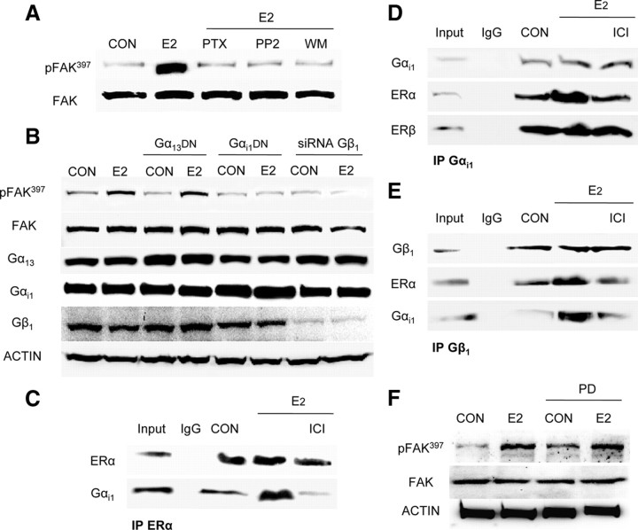
Fig. 3.
ERα signaling to FAK requires Gαi/Gβ. A, Breast cancer cells were exposed for 20 min to 10 nm E2, in the presence or absence of the G protein inhibitor PTX (100 ng/ml), of the c-Src inhibitor, PP2 (0.2 μm), or of the PI3K inhibitor WM (30 nm), and Tyr397 FAK phosphorylation was assayed with Western analysis. B, Breast cancer cells were treated with E2 (10 nm) after transfection with dominant-negative Gα13 or Gαi constructs or siRNAs vs. Gβ1. Gα13, Gαi, Gβ1, actin, FAK, or phospho-FAK397 was assayed in cell extracts. C–E, T47-D cell protein extracts were immunoprecipitated with antibodies toward ERα, Gαi1, or Gβ1, and coimmunoprecipitation of Gαi1 (C), ERα and ERβ (D), and ERα and Gαi1 (E) was tested by Western analysis. F, Breast cancer cells were exposed for 20 min to 10 nm E2, in the presence or absence of the MAPK inhibitor PD98059 (PD; 5 mm), and Tyr397 FAK phosphorylation was assayed with Western analysis. CON, Control; IP, Immunoprecipitation. All experiments were performed three times, and representative blots are presented.

