
Fig. 4.
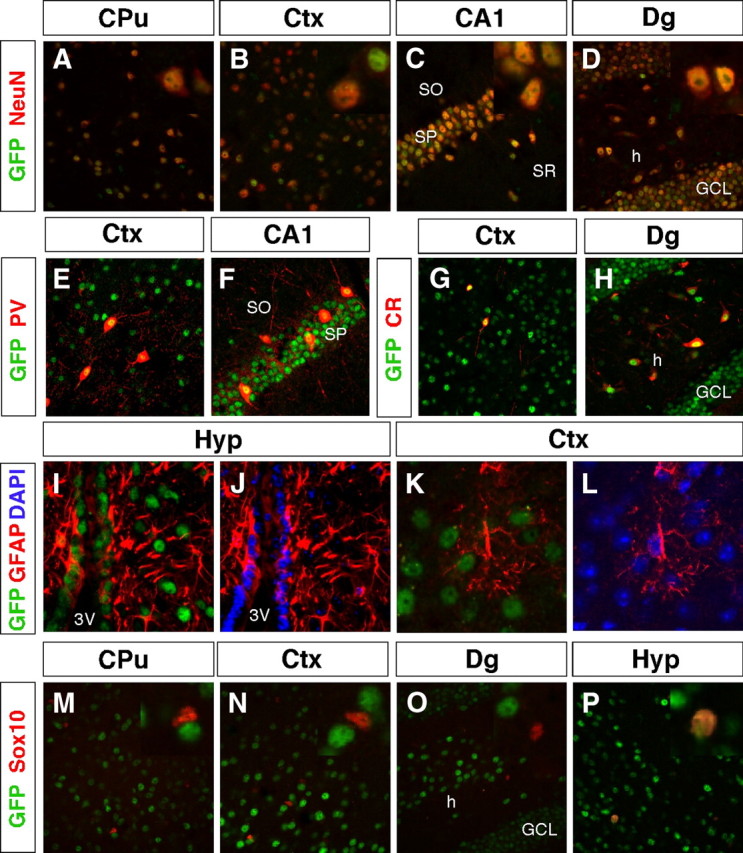
TRα1-GFP is expressed in virtually all neurons but in only a few specialized glial cells. A–P, Confocal imaging micrographs were taken of adult sections of the brain after immunohistochemistry against GFP to detect TRα1-GFP and markers for different cell types. Insets show high magnifications of the same region. A–D, TRα1-GFP expression was detected in all analyzed neurons (NeuN+ cells) in the adult brain, including in the caudate putamen (A), somatosensory cortex (B), CA1 region (C), and the dentate gyrus (D). E–H, Expression in GABAergic interneurons. TRα1-GFP was detected in PV+ interneurons in the cortex (E) and in the CA1 region of the hippocampus (F). TRα1-GFP and CR were coexpressed in interneurons of the cortex (G) and in interneurons and mossy cells in the hilus of the hippocampus (H). I–L, TRα1-GFP expression was detected in GFAP+ tanycytes lining the third ventricle of the hypothalamus (I and J), but not in astrocytes as shown in the cortex (K and L). Nuclei were counterstained with 4′6,-diamidino-2-phenylindole (DAPI). M–P, Double immunolabeling of oligodendrocytes with Sox10 show that TRα1-GFP was expressed only in adult oligodendrocytes of the hypothalamus (P), but not in the caudate putamen, cortex, or dentate gyrus (M–O). CPu, Caudate putamen; Ctx, cortex; GCL, granular cell layer; h, hilus; Hyp, hypothalamus; SO, stratum oriens; Sox, SRY related high-mobility group box; SP, stratum pyramidale; SR, stratum radiatum; 3V, third ventricle.