
Fig. 5.
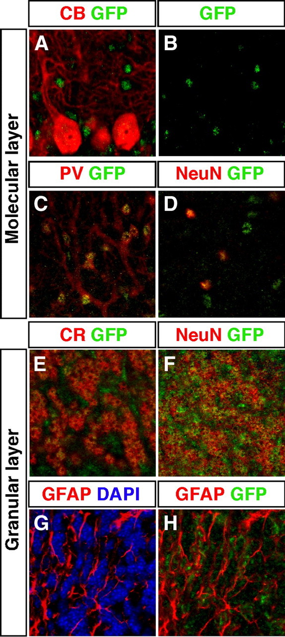
TRα1-GFP is expressed in specific neurons and glial cells in the cerebellum. A–H, Confocal imaging micrographs of adult sections showing expression of TRα1-GFP (detected with GFP antibody) and markers for different cell types. Examination of Purkinje cells marked with calbindin (CB) showed no colocalization with TRα1-GFP (A and B). In contrast, in the molecular layer, TRα1-GFP was expressed in PV+ stellate/basket cells (C) and in all neurons marked with NeuN (D). In the granular layer TRα1-GFP was expressed in granular cells as shown by immunostaining for both CR (E) and NeuN (F). TRα1-GFP was also expressed in GFAP+ glia cells (G and H). Nuclei were counterstained with 4′6,-diamidino-2-phenylindole (DAPI) in G.