Fig. 1.
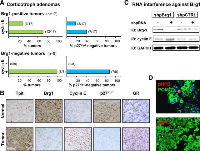
Cyclin E up-regulation and loss of p27Kip1 expression in corticotroph adenomas. A, A panel of 25 human corticotroph adenomas was studied by immunohistochemistry for Brg1, cyclin E, and p27Kip1 expression. All samples were also positive for ACTH, Tpit, and GR expression. Normal pituitary tissue is always positive for nuclear Brg1 and p27Kip1 and negative for cyclin E. The upper panels represent data for adenomas that are Wt for Brg1 (i.e. positive) whereas the lower panels represent data for the Brg1-negative subset. For each, the left panel indicates percentage of tumors that are either cyclin E negative (Wt) or cyclin E positive, and the right panels indicate the proportion of each tumor group that has lost expression of p27Kip1. No tumor was ever observed to be Brg1 negative and cyclin E negative. B, Immunohistochemical analyses for a representative human Brg1-negative, cyclin E-positive, p27Kip1-negative adenoma. Normal pituitary is shown for comparison. C, The putative repressor activity of Brg1 on cyclin E expression was verified in AtT-20 cells using RNA interference with a short hairpin loop RNA target against Brg1 (shpBrg1) in comparison with a scrambled-sequence control RNA (shpCTRL). Western blot analysis of Brg1, cyclin E, and GAPDH indicated successful knockdown of Brg1 resulting in up-regulation of cyclin E. D, Sections from two representative tumors of the Brg1-negative, cyclin E-positive, and p27Kip1-negative group showing colabeling of POMC-positive cells with pHH3. All tumor samples of this group showed similar double-positive cells, but no pHH3-positive, ACTH-negative cells. IB, Immunoblotting.

