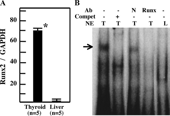
Fig. 3.
Expression of Runx2 in normal thyroid glands. A, Quantitative PCR for Runx2 in thyroid gland (n = 5) and liver (n = 5) from 12-wk-old wild-type mice. Quantitative PCR was carried out as described in the Materials and Methods. Means of Runx2/GAPDH ± sd are indicated. *, P < 0.001. B, EMSA with Oligo OC. Nuclear extracts (NE) from thyroid gland (T) and liver (L) from wild-type mice were incubated with radiolabeled Oligo OC, which corresponds to the Runx2 binding site on the osteocalcin gene promoter. Protein/DNA complexes are indicated by black arrows. Complex formation was inhibited by addition of self-competitor (compet) and diminished by incubation with anti-Runx2 antibody (Runx) (1:250) but not with control serum (N). Arrow indicates Runx2/Oligo complex. Ab, Antibody.

