
Fig. 5.
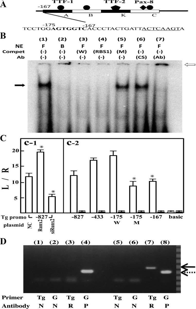
Runx2 binding to Tg promoter region. Panel A, Scheme of Tg promoter structure and 5′-upstream sequence from region A. Rat Tg gene promoter (from −167 to −1 bp; transcription start site is designated as +1), which is essential for thyroid-specific expression, consists of regions A, B, K, and C. Runx2 binding consensus sequence, AGTGGTC, shown in bold, is located just upstream from region A. The underlined sequence is the TTF-1 binding site in region A. Panel B, EMSA with Oligo W. Nuclear extracts (NE) from FRTL-5 cells (F) and BRL-3A liver cells (B) were incubated with radiolabeled Oligo W, which corresponds to −178 to −167 bp of rat Tg promoter. Protein/DNA complex is indicated by black arrows. Complex formation was inhibited by the addition of self-competitor (W) and Oligo RBS1 containing Runx2 binding consensus sequences but not by Oligo M (M) to which mutations were introduced (lanes 3–5). In lanes 6 and 7, radiolabeled Oligo W was incubated with control serum (CS) (lane 6) or anti-Runx2 antibody (Ab) (1:250, lane 7). The supershifted band is indicated by a white arrow. Panel C, Promoter activity of rat Tg-luciferase chimera plasmids. C-1, pTg-Luc (−827) (2 μg) was cotransfected with 2 μg of pSilencer NC, pcDNA3-Runx2 (Runx2), or pSilencer-siRunx2 (siRunx2) into FRTL-5 cells by electroporation. Data are means of triplicate assays. *, P < 0.01 vs. NC. C-2, Luciferase activities in lysates from FRTL-5 cells (white bars) and BRL-3A cells (black bars) after transfection with pTg-Luc (−827) and its deletion mutants, as indicated. Data are means of triplicate assays. pTg-Luc (−175M) has a mutation in the Runx-2 binding site. *, P < 0.01 vs. pTg-Luc(−175W). All cells were cotransfected with 100 ng pRL-SV40 to normalize transfection efficiency. Data are means ± sd. Panel D, ChiP assays of thyroid gland material. Liver (lanes 1–4) and thyroid glands (lanes 5-8) were cross-linked with 1% formaldehyde and then precipitated with anti-Runx2 antibody (2.5 μg, R), anti-RNA polymerase II antibody [2.5 μg, positive control (P)], and normal mouse IgG [1 μg, NC (N)]. Precipitated DNA fragments were amplified using primers for Tg promoter or for GAPDH promoter (G). Solid arrow indicates Tg promoter cDNA, and dotted arrow indicates GAPDH promoter cDNA.