
Fig. 1.
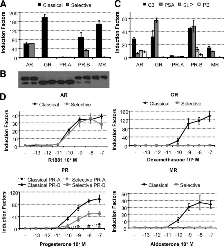
Comparison of receptor selectivity of classical and selective enhancers and promoters. A, HEK293 cells (104 per 96 wells) were transfected with 100 ng of a classical or a selective ARE-based reporter and 10 ng of a receptor expression vector (see Table 4 for the ARE sequences). Cells were stimulated for 24 h with R1881, dexamethasone, progesterone, or aldosterone (all at 10−8 m). Results are presented as induction factors. Error bars are the averages’ sem of at least three independent experiments performed in triplicate. B, For immunoblotting, transfected HEK293 cells were treated for 1 h without or with 10 nm of the relevant hormone. The expressed proteins were detected using an anti-Flag antibody. C, HEK293 cells were transfected as in A with 100 ng of a reporter gene driven by a single copy of the classical C3(1 )-enhancer and PSA-promoter or the selective slpARU-TK-TATA and probasin promoter. Cells were stimulated for 24 h with 10 nm R1881, dexamethasone, progesterone, or aldosterone. Results are presented as in A. D, HEK293 cell lines containing a stable integrated classical or selective ARE were made using the Flp-In T-REx system (Invitrogen). The indicated receptor expression vector (100 ng) was transiently transfected. Cells were stimulated for 24 h with different concentrations of R1881, dexamethasone, progesterone, or aldosterone. Results are presented as in A.