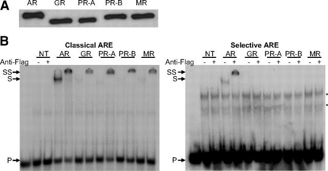
Fig. 2.
EMSA of nonselective and selective AREs by full-size receptors. A, COS-7 cells were transfected with 8 μg receptor expression plasmid. Cells were treated for 1 h with 10 nm R1881, dexamethasone, progesterone, or aldosterone. Nuclear extracts were made as described in Materials and Methods. The expressed proteins were blotted and detected using the M2 anti-Flag antibody. B, 32P-labeled classical (SLP-MUT) or selective (SLP-HRE) probes were incubated with similar amounts of nuclear extract from nontransfected (NT) COS-7 cells or nuclear extracts containing the AR, GR, PR-A, PR-B, or MR. No protein was added in the first lane as a negative control. Anti-Flag antibody was added as indicated at the top. Arrows indicate the positions of the unbound (P), shifted (S), and supershifted (SS) probe. Asterisks indicate nonspecific complexes.

