
Fig. 2.
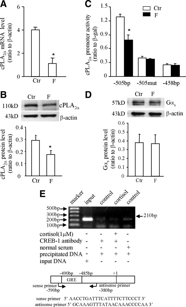
A and B, Inhibition of cPLA2α mRNA and protein expression by cortisol (F, 1 μm) in HFL-1 cells. The top panel of B is the representative immunoblot; bottom panel shows the mean data. C, Inhibition of cPLA2α promoter (−505 bp) activity by cortisol (F, 1 μm) could be diminished either by introduction of triple-nucleotide mutations (−505mut) into the putative GRE at −485 to −490 bp or by complete removal of the GRE as −458 bp in HFL-1 cells. D, Cortisol (F, 1 μm) treatment did not affect the expression of Gαs protein in HFL-1 cells. The top panel of D is the representative immunoblot; bottom panel shows the mean data. n = 3–4. *, P < 0.05 vs. control (Ctr). E, ChIP demonstrated that cortisol (1 μm) caused no obvious binding of CREB to the GRE in the cPLA2α promoter as revealed by gel electrophoresis of the PCR products amplified from the DNA fragments precipitated by CREB antibody in HFL-1 cells. The top panel is a representative gel of three individual experiments; bottom panel illustrates the positions and sequences of the primers used for PCR.