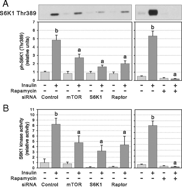
Fig. 2.
S6K1 activation after RNA interference or chronic rapamycin treatment of 3T3-L1 adipocytes. Cells were electroporated with control or target siRNAs or treated (24 h) with rapamycin (25 nm) or DMSO and serum deprived before 45 min stimulation with 10 nm insulin. A, Western blotting of cell lysates was performed with phosphospecific antibody against S6K1 Thr389 (n = 5). B, S6K1 kinase activity was measured using S6 peptide (RRRLSSLRA) as substrate (n = 3). Densitometry measures of phosphorylation and kinase activity were normalized for the expression of S6K1 and for protein content, respectively. Results in panel A show representative gels and densitometric analysis. Results are shown as means ± se. Groups with different letters are significantly different (P < 0.05).

