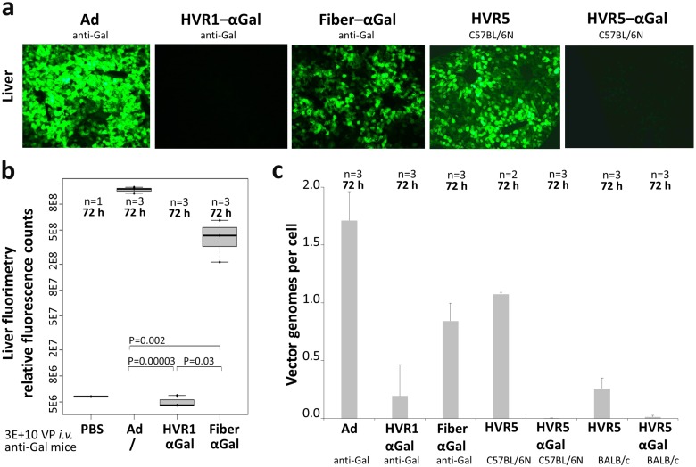
Fig 8. Ad–αGal immunocomplexes altered in vivo transduction of liver after systemic delivery of vectors into anti-Gal mice.
Transgene product expression was analyzed at 72 h after i.v. delivery of 3E+10 physical vector particles of EGFP-expressing vectors and differed significantly between vectors decorated with αGal at distinct capsomers. (a) Fluorescence microscopy of liver cryosections depicts transduction results of Ad control vector, HVR1–αGal AICs, and Fiber–αGal AICs in anti-Gal mice, and Ad HVR5 and Ad HVR5–αGal in αGal-expressing C57BL/6N mice. (b) Fluorimetry of anti-Gal mouse livers quantified transgene expression by Ad control vector, or HVR1–αGal or Fiber–αGal AICs. (c) Quantitative PCR detected vector genomes delivered into livers of anti-Gal mice (Ad, HVR1–αGal AICs, Fiber–αGal AICs), and αGal-expressing C57BL/6N mice and BALB/c mice (Ad HVR5, Ad HVR5–αGal). P values were calculated by unpaired, two-tailed t-test assuming equal variances.

