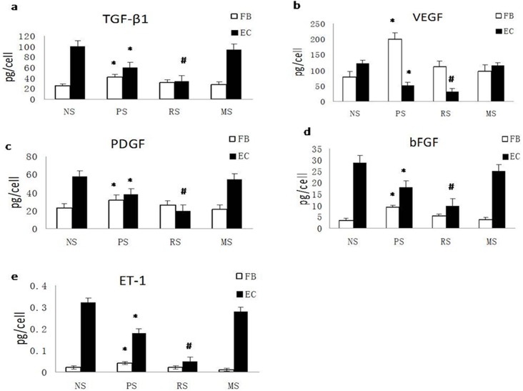
Fig 3. TGF-β1, VEGF, PDGF, ET-1, and bFGF secretion is suppressed in ECs but increased in fibroblasts during scar development.
A. Quantification of the secretion of TGF-β1 (a), VEGF (b), PDGF (c), bFGF (d), and ET-1 (e) from fibroblasts and ECs from NS, PS, RS and MS. NS, PS, RS and MS represent normal skin, proliferative scar, regressive scar and mature scar, respectively. * P<0.05, # P<0.01, n = 8.

