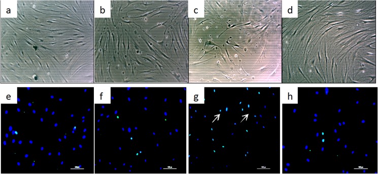
Fig 5. EC-conditioned media induce cell apoptosis.
The morphology of fibroblasts was imaged under a light microscope after culture with NS (a), PS (b), RS (c) and MS EC-conditioned media (d). Fibroblast apoptosis was viewed using immunofluorescence staining after co-culture with NS (e), PS (f), RS (g) and MS EC-conditioned media (h). Apoptotic nuclei were stained green and are indicated by arrows, whereas negative nuclei were stained blue. NS, PS, RS and MS represent normal skin, proliferative scar, regressive scar and mature scar, respectively. The scale bar represents 50 μm, n = 5.

