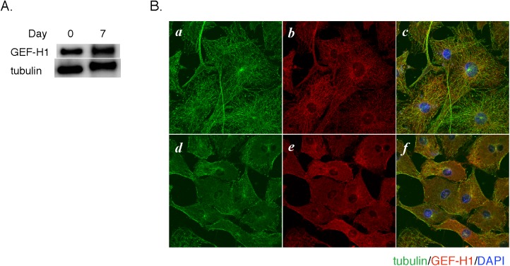
Fig 3. Expression and localization of GEF-H1.
A. Cell lysates from 3T3-L1 preadipocytes (Day 0) or differentiated adipocytes (Day 7) were subjected to SDS-PAGE and immunoblotting with anti-GEF-H1 and anti-β-tubulin antibodies. B. 3T3-L1 preadipocytes grown on a cover glass were treated without (a-c) or with (d-f) sucralose (20 mM) for 2 hours. After fixation with 100% methanol at -20°C for 2 minutes, cells were immunostained for β-tubulin (green, a and d) and GEF-H1 (red, b and e). Cell nuclei were visualized with DAPI (blue) in the merged images (c and f).

