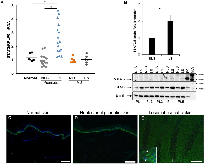
Fig 1. STAT2 expression is elevated in psoriatic skin.
(A) STAT2 mRNA expression was examined by qPCR in biopsies obtained from normal healthy volunteers as well as from paired punch biopsies obtained from nonlesional (NLS) and lesional (LS) skin from patients suffering from psoriasis or atopic dermatitis (AD). The mRNA expression of RPLP0 was used for normalization. Biopsies from 6 healthy volunteers, 16 psoriatic patients and 6 atopic dermatitis patients were examined. The results are presented as dot plots with the horizontal line expressing the mean value. All measurements were performed in triplicates. (B) Whole cell protein extracts were prepared from paired keratome biopsies taken from nonlesional and lesional psoriatic skin from five psoriatic patients. Phosphorylated STAT2 as well as total STAT2 protein was analyzed by western blotting. Equal protein loading was assessed by detecting the protein level of β-actin. Protein extract from keratinocytes stimulated with IFNα for 1 hour was included as a positive control (PC). MW; molecular weight marker. Densitometic analysis of the band intensity was carried out and values were normalized to β-actin. *P < 0.01. (C-E) Immunofluorescence analysis was performed on paraffin-embedded punch biopsies from (C) normal skin as well as (D) nonlesional and (E) lesional psoriatic skin. Nuclear staining was performed using 4’, 6-diamidine-2’-phenylindole dihydrochloride (DAPI). Green color (Alexa Fluor 488) represents STAT2 protein. Three sets of biopsies from three different patients were investigated. Arrows show nuclear staining. Scale bar = 100 μm.

