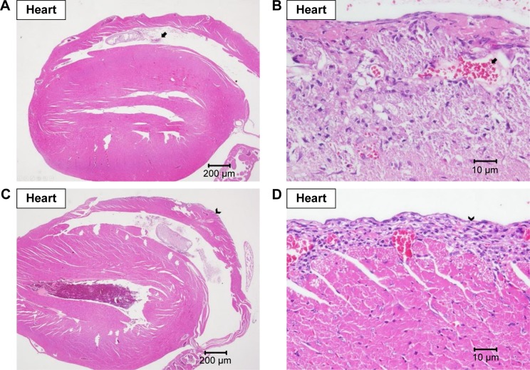
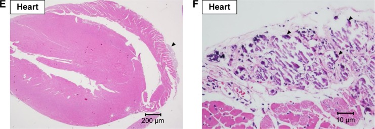
Figure 5.
Histopathological analysis of heart.
Note: Histopathological analysis with H&E staining of heart at (A) 20× and (B) 400× magnifications with focal fibrotic emboli (arrow), at (C) 20× and (D) 400× magnifications with focal fibrosis (arrow head), and at (E) 20× and (F) 400× magnifications with epicardial mineral deposition (triangle).


