Figure 1.
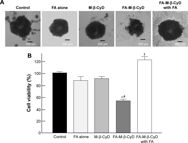
Cytotoxic activity of M-β-CyDs for spheroids of KB cells.
Notes: (A) Macroscopic observation of spheroids of KB cells. (B) Cell viability assay by MTT assay. KB cells were cultured for 72 h with RPMI medium (FA (−)) and 10× spheroid formation ECM at 37°C. KB cells were treated with M-β-CyDs (5 mM) for 24 h at 37°C in the presence or absence of 1 mM FA. After washing twice with RPMI medium, MTT assay was performed. Bar graphs represent mean ± SEM (n=3–4 per group). Significant difference with P<0.05 as compared to *control, #M-β-CyD, and ‡FA-M-β-CyD.
Abbreviations: ECM, extracellular matrix; FA, folic acid; MTT, 3-(4,5-dimethylthiazol-2-yl)-2,5-diphenyltetrazolium bromide; RPMI, Roswell Park Memorial Institute; SEM, standard error of the mean.

