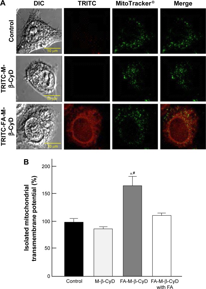
Figure 2.
Colocalization of M-β-CyDs with mitochondria and its effects on mitochondrial membrane potential.
Notes: (A) Colocalization of TRITC-M-β-CyDs with mitochondria. KB cells were treated with 10 µM TRITC-M-β-CyDs for 2 h, and then the cells were treated with MitoTracker. The cells were then scanned with a confocal laser microscope. The representative images are shown (n=3 per group). (B) Effects of M-β-CyDs on mitochondrial transmembrane potential of isolated mitochondria from KB cells (FR-α (+)). The isolated mitochondria were incubated with 50 µM M-β-CyDs for 1 h in the presence or absence of 500 µM FA. A mitochondrial transmembrane potential was measured by rhodamine 123 staining with a plate reader. Bar graphs represent mean ± SEM (n=3–6 per group). Significant difference with P<0.05 as compared to *control and #M-β-CyD.
Abbreviations: DIC, differential interference contrast; FA, folic acid; FR, folate receptor; SEM, standard error of the mean; TRITC, tetramethylrhodamine isothiocyanate.

