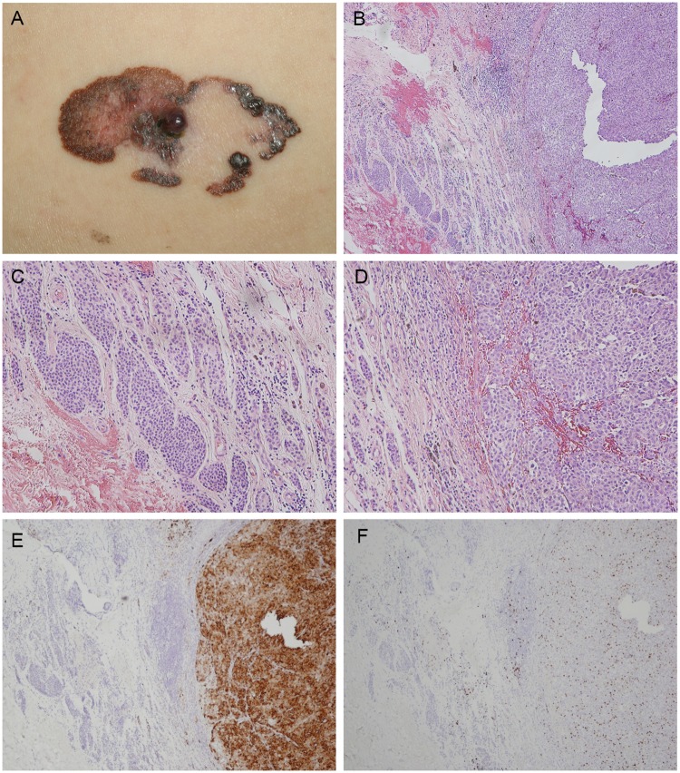
Fig 1. Superficial spreading melanoma arising from a nevus.
(A) A 63-year-old man presented with a 5 x 2.5 cm plaque on the left flank with irregular scalloped borders, mottled variegate color, large areas of regression, and a small nodule in the center. (B) Low-powered photomicrograph demonstrating a nodular lesion composed of tumor cells arranged in nests in the dermis with a Breslow thickness of 3 mm. Mitotic figures are numerous. Original magnification x 40. (C) Conventional dermal nevus nests can be noted adjacent to the tumor mass. Original magnification x 100. (D) High-powered photomicrograph reveals nests of tumor cells with vesicular nuclei and prominent nucleoli. Original magnification x 100. (E) Melanoma cells stained positive and nevus cells negative for HMB-45. Original magnification x 40. (F) Melanoma cells stained positive for Ki-67. Original magnification x 40.

