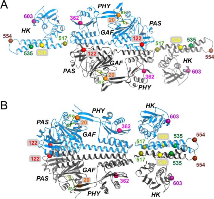
Figure 1.

Locations of spin-label sites in the Agp1 phytochrome. A, crystal structure of the Agp1-PCM in antiparallel arrangement (PDB code 5HSQ) and two monomers of a homology model for the Agp1 His kinase (template from a T. maritima His-kinase (12)) placed manually close to the C termini of each PCM module. B, crystal structure of the Agp1-PCM in parallel arrangement (PDB code 5I5L) and a homology model for the Agp1 His-kinase dimer (12) placed manually next to the C termini of the PCM modules. The Cα atoms of the residues mutated to cysteines, as well as of the functional Cys-20, are shown as pairwise colored spheres and labeled according to their position in Agp1.
