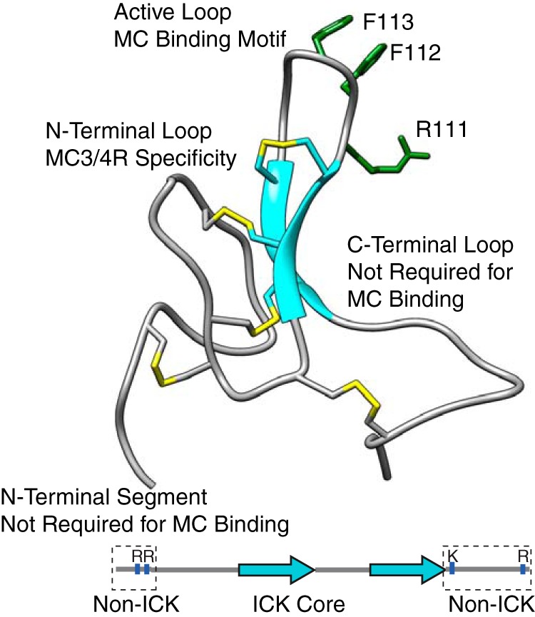
Figure 1.

AgRP (83–132) NMR structure (PDB code 1HYK) and schematic indicating ICK and non-ICK regions. The structure of AgRP includes the functional domains shown here and their contribution to MC4R binding. The disulfide bonds are shown in yellow. The schematic highlights the N-terminal segment and C-terminal loop, which are conserved in mammalian sequences. The active loop possesses an RFF triplet (residues 111–113) necessary for melanocortin receptor binding. Positively charged residues within these non-ICK domains are indicated.
