Figure 1.
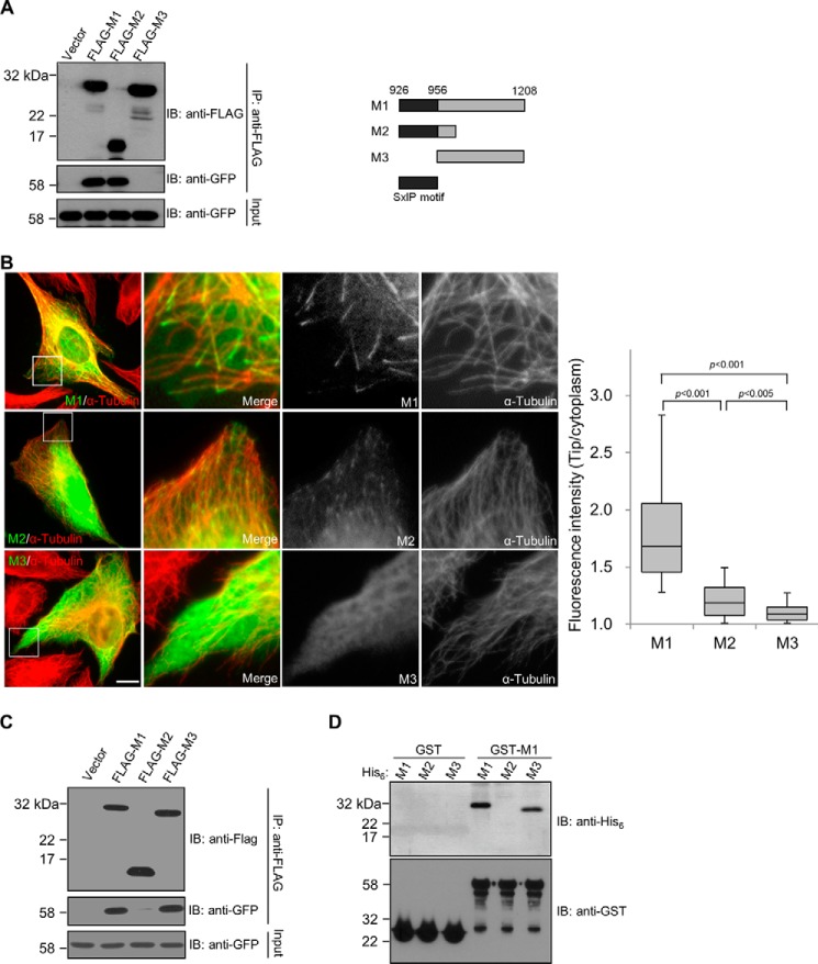
The plus-end-tracking domain of CDK5RAP2 contains an SXIP motif and a dimerization domain. A, EB1 binding of CDK5RAP2 fragments were tested by co-immunoprecipitation (IP) from the extracts of HEK293T cells co-expressing the CDK5RAP2 fragments (FLAG tagged) and EB1-GFP. The anti-FLAG immunoprecipitates (30%) and the cell extract inputs (5%) were analyzed by immunoblotting (IB). Shown on the right is a scheme of the CDK5RAP2 fragments. B, GFP-tagged M1, M2, and M3 were transiently expressed in U2OS cells. We analyzed cells expressing the proteins at similar levels and measured the fluorescence intensities at the microtubule distal tips and in the cytoplasm. Boxed areas are enlarged. Bar, 10 μm. The tip/cytoplasm intensity ratios of 50 microtubule tips selected from 5 transfected cells are presented as box-and-whisker plots: boxes represent the 25th and 75th percentiles, a line within the boxes depicts the median, and whiskers represent the 10th and 90th percentiles. p values were calculated by the Student's unpaired two tails t test. C, GFP-M1 was cotransfected with FLAG-tagged M1, M2, or M3 into HEK293T cells. After anti-FLAG immunoprecipitation (IP), the immunoprecipitates (30%) and the cell extract inputs (5%) were analyzed on immunoblots (IB). GFP-M1 was co-immunoprecipitated with FLAG-tagged M1 and M3, but not with FLAG-M2. D, an in vitro binding assay was conducted with 0.5 μm, each of the purified recombinant proteins His6-M1, His6-M2, His6-M3, GST, and GST-M1. After incubation of the His6-tagged proteins with the GST proteins as indicated, the GST proteins were pulled down, and the bound proteins were analyzed on anti-His6 and anti-GST immunoblots (IB). His6-tagged M1 and M3 were specifically found in the GST-M1 pulldowns, whereas His6-M2 was not detected.

