Figure 6.
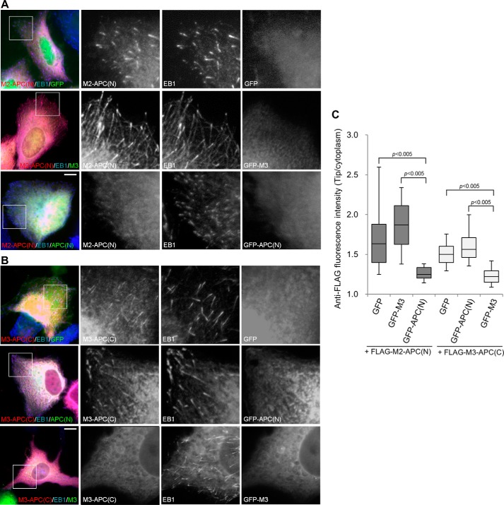
Disruption of SXIP dimerization interferes with microtubule plus-end tracking. A, U2OS cells were transiently cotransfected with plasmids encoding FLAG-M2-APC(N) and a GFP fusion protein (GFP, GFP-M3, or GFP-APC(N)). The cells were stained with anti-FLAG and anti-EB1 antibodies. B, FLAG-M3-APC(C) was transiently co-expressed with GFP, GFP-APC(N), or GFP-M3 in U2OS cells. A and B, boxed areas are enlarged. Bars, 10 μm. C, the intensities of anti-FLAG immunofluorescence were measured at microtubule tips as labeled by anti-EB1 staining and in the cytoplasm. In each group, 80–100 microtubule tips were measured from 8–10 cells that expressed the FLAG-tagged proteins and the GFP proteins at low and moderate levels, respectively. In the box-and-whisker plots, boxes represent the 25th and 75th percentiles, a line within the boxes depicts the median, and whiskers represent the 10th and 90th percentiles. The significance of differences between the indicated values was evaluated by Student's unpaired two-tailed t test.

