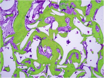
Fig. 1.

Histomicrograph illustrating the various tissue areas measured on the sections: newly formed bone (green mask), soft tissues (purple mask), and “others”, including residual bone substitute particles and empty spaces either due to removal of the bone substitute particles during to the decalcification processing or due to artifacts (white mask)
