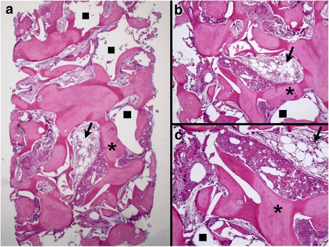
Fig. 2.

Histomicrograph of a biopsy from the BC group. a Overview—×25 magnification; b ×30 magnification; c ×60 magnification. Areas corresponding to BC removed during histological processing (square) in direct contact with newly formed bone (asterisk), containing a large number of osteocytes, and with soft tissue (arrow) can be observed (hematoxylin-eosin stain)
