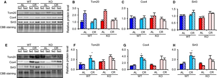
Figure 3.

Srebp‐1c was required for CR‐associated upregulation of proteins involved in mitochondrial biogenesis in WAT but not in the liver. (A–D) Liver and (E–H) WAT. Example of immunoblot images showing expression of proteins involved in mitochondrial biogenesis in liver tissue (A) and WAT (E) samples from eight groups of mice (WTAL‐fed, WTAL‐fast, WTCR‐fed, WTCR‐fast, KOAL‐fed, KOAL‐fast, KOCR‐fed, KOCR‐fast). Quantitative analysis of immunoblots was performed using a chemiluminescence method. Results for Tom20 (B, F), Cox4 (C, G), and Sirt3 (D, H) are each expressed as relative intensity of the indicated protein/CBB staining compared with values in the WTAL‐fed group (n = 4 per group). Values in all panels are means ± SEM. *P < 0.05 vs. AL, analyzed by Tukey's test.
