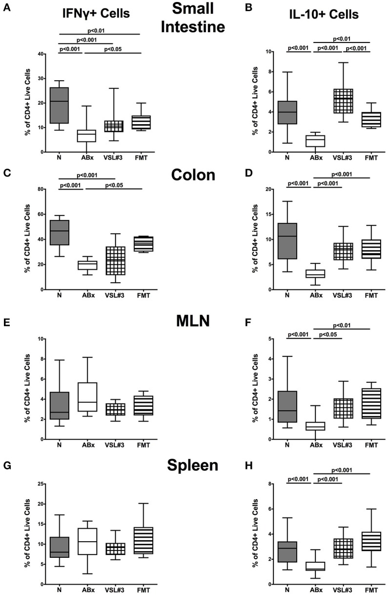
Figure 9.

IFN-γ and IL-10 producing CD4+ cells in intestinal and systemic compartments of secondary abiotic mice following recolonization with VSL#3 or complex murine microbiota. Lymphocytes were isolated from small intestinal and colonic lamina propria, MLN, and spleen and stimulated with PMA/ionomycin in presence of brefeldin A and subsequently analyzed by flow cytometry. The percentages of IFN-γ (left panel A,C,E,G) and IL-10 (right panel B,D,F,H) producing CD4+ cells in the small intestine (A,B), colon (C,D), MLN (E,F), and spleen (G,H) in naive conventional mice (N), by antibiotic treatment generated secondary abiotic mice (ABx), and mice subjected to VSL#3 recolonization or fecal microbiota transplantation (FMT) were determined on day 28 following peroral reassociation. Box plots represent the 75th and 25th percentiles of the medians (black bar inside the boxes). Total range and significance levels (p-values) determined with one-way ANOVA test followed by Tukey post-correction test for multiple comparisons are indicated. Data shown were pooled from two independent experiments (n = 10–15/group).
